
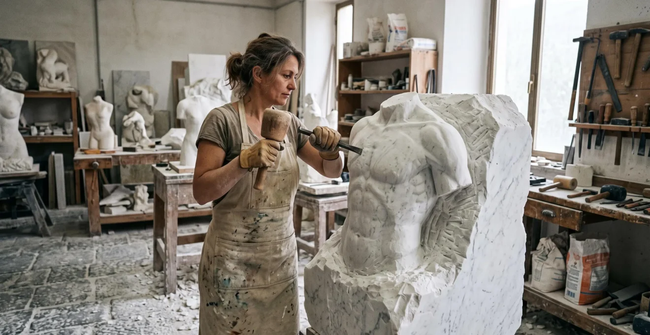
Michelangelo did not need a medical degree to understand the human body with extraordinary sophistication. His anatomical knowledge developed through direct observation, artistic apprenticeship, access to human dissections, and sustained investigation of how bones, muscles, and skin interact in movement.
Historical accounts describe his examination of human cadavers at Santo Spirito in Florence. That experience complemented a workshop education centered on drawing, modeling, and studying visible form. Rather than memorizing isolated muscle shapes, he learned to understand the relationships that make a figure structurally convincing.
His sculptures were not flawless anatomical diagrams. Michelangelo frequently enlarged, compressed, or emphasized features to communicate strength, tension, spirituality, or grief. His achievement lay in knowing enough about the body’s construction to make such departures persuasive.
For contemporary figurative sculptors and realist painters, the practical lesson is that anatomical accuracy develops through a combination of structural knowledge, observation, three-dimensional reasoning, and purposeful artistic decisions.
Michelangelo’s anatomical method in three principles
- Study beneath the surface: Understanding bones, muscle attachments, and movement produces more reliable figures than copying visible contours alone.
- Construct living tissue: Convincing anatomy depends on skin, fat, tension, and transitions between forms—not merely displaying individual muscles.
- Use accuracy selectively: Structural knowledge establishes credibility, while deliberate departures from anatomical proportion can strengthen expression.
- Why Did Leonardo Dissect 30 Corpses to Paint More Convincing Human Figures?
- How Do You Sculpt Convincing Skin When You Can Only See Bones Beneath?
- Academic Proportion Systems vs Life Observation: Which Produces More Convincing Figures?
- The Insertion Point Mistake: Why Your Perfectly Muscled Figures Still Look Incorrect
- The Accuracy Decision: When to Sacrifice Correct Anatomy for Emotional Impact
- How Do You See a Finished Sculpture Inside a Raw Quarried Block Before You Start?
- Online Tutorials vs In-Person Mentorship: Which Builds Genuine Classical Painting Skills?
- How Do Sculptors Reveal Fluid Human Forms Locked Inside Rigid Marble Blocks?
Why Did Leonardo Dissect 30 Corpses to Paint More Convincing Human Figures?
Leonardo da Vinci dissected human bodies to understand the structures responsible for visible form, movement, and bodily function. The Royal Collection Trust estimates that he examined perhaps thirty human corpses, but this is not an independently verified total. His investigations extended far beyond improving paintings and included extensive scientific inquiry into human anatomy.
Renaissance artists faced a fundamental problem: observing the outside of the body reveals where a bulge appears, but not necessarily why it appears. A raised shoulder might result from skeletal position, muscle contraction, or the movement of surrounding tissue. Without understanding these causes, an artist could reproduce one pose convincingly yet struggle to construct another.
Dissection provided a way to investigate underlying structures rather than relying exclusively on surface imitation. According to the Royal Collection Trust’s study of Leonardo’s anatomical research, his drawings examined successive anatomical layers and multiple viewpoints, particularly during his intensive investigations of bones and muscles in the early sixteenth century.
Perhaps 30 human corpses
The Royal Collection Trust’s qualified estimate of Leonardo’s dissections. The figure should not be interpreted as an exact count or as bodies examined exclusively for painting.
Royal Collection Trust, Leonardo’s Study of Anatomy.
For figurative artists, three interconnected levels of anatomical understanding are especially useful:
- Skeletal framework: The bones and joints establish proportions, stable landmarks, movement limits, and the underlying architecture of a pose.
- Muscle origins and insertions: Attachment relationships explain how muscular masses change direction, length, and prominence during movement.
- Superficial anatomy: Muscle, tendon, fat, fascia, and skin determine which structural features actually appear on the body’s surface.
Michelangelo pursued related knowledge through a different artistic career. Historical research published in the Journal of Anatomy review on anatomy and Renaissance art education describes his access to human dissection at Santo Spirito in Florence as early as 1493. Accounts connect this opportunity with his relationship to the religious community and his work on a wooden crucifix.
These details come partly through historical biography, including testimony associated with Ascanio Condivi, rather than a complete record of every anatomical session. The evidence supports participation in dissection, not the claim that Michelangelo acquired perfect knowledge of every human structure.
Nor should Leonardo’s achievements be transferred wholesale to Michelangelo. Leonardo investigated circulation, nerves, organs, and reproduction as well as artistic anatomy. Michelangelo’s surviving artistic work and biographical record instead demonstrate a particularly strong concern with human form, muscular organization, and expressive movement.
Neither artist needed the institutional qualifications associated with modern medical training to investigate anatomy. Workshops, direct observation, access to cadavers, and extensive drawing practice offered another demanding educational route.
Dissection explains how an artist could discover the body’s hidden architecture. The next challenge is translating that architecture into the visible appearance of living flesh.
How Do You Sculpt Convincing Skin When You Can Only See Bones Beneath?
Convincing sculpted skin begins with fixed skeletal landmarks, then builds outward through muscular volume, fat distribution, and surface tension. Bones establish reliable positional references, but skin does not simply follow their contours. The sculptor must interpret how tissue stretches, compresses, and moves across the underlying structure in a particular pose.
The clavicles, sternum, rib cage, iliac crests, kneecaps, and prominent joints provide useful reference points because their positions are tied closely to the skeleton. Their visibility changes with body composition, posture, and lighting, but their structural relationships remain useful guides.
A practical construction process begins by locating these landmarks before developing the larger muscular masses. On a clay torso, for example, the rib cage and pelvis can be established first. The pectoral region, abdominal wall, and oblique muscles can then be organized around those volumes rather than applied as disconnected surface decorations.
Bone-to-surface construction becomes especially important when the figure twists. The apparent shape of a muscle changes with perspective and joint position, even though its underlying attachments remain anatomically related.
Renaissance artists could deepen this understanding through observation of living models, anatomical drawings, and studies of flayed figures known as écorchés. Such models expose muscular organization without requiring direct access to a human dissection.
Once the primary forms are established, the sculptor must read muscular tension. A supporting leg, raised arm, or tightly flexed hand creates a particular distribution of mechanical effort. Some tissues become prominent while others remain comparatively soft.
Imagine a standing figure shifting its weight onto the right leg. The pelvis and supporting hip must accommodate the transfer of load. If every muscle on both legs receives identical definition, the sculpture may look symmetrical and rigid despite an otherwise convincing pose.
The test is not whether each muscle is recognizable. It is whether the visible distribution of tension is compatible with the depicted action.
Skin and superficial fat are equally important. Their thickness varies across the body and affects the visibility of muscles, tendons, veins, and bony landmarks. A soft transition across the abdomen can communicate living tissue more effectively than deeply incised boundaries between every abdominal muscle.
Michelangelo’s treatment of anatomical surfaces was often idealized and emphatic, but his figures demonstrate the expressive possibilities of continuous transitions between major masses.
For modern sculptors, a useful exercise is to model the same limb in a relaxed position and then under muscular tension. Compare the silhouette, compression around the joint, and prominence of tendons before adding fine surface detail.
Convincing skin emerges from relationships between structural layers. Once those relationships are understood, the next question is whether proportion should follow a predetermined canon or the actual person being observed.
Academic Proportion Systems vs Life Observation: Which Produces More Convincing Figures?
Neither academic proportion systems nor life observation is universally superior. Proportion systems provide repeatable construction rules, especially when working from imagination. Life observation reveals individual variation, weight, asymmetry, and gesture. The most convincing figurative work often combines the two: measured structure establishes coherence, while observation prevents the figure from becoming mechanically standardized.
Renaissance artists inherited ideals associated with classical proportion, including ideas attributed to Vitruvius. Later artists such as Albrecht Dürer investigated bodily measurement through systematic approaches of their own.
These methods attempt to make the complex human figure manageable by relating parts to a reference unit. The head, for example, can serve as a convenient comparative measure when constructing an entire standing figure.
Such systems are useful, but a proportional canon is not a universal anatomical law. Actual bodies differ in head size, limb length, shoulder breadth, pelvic structure, age-related characteristics, and overall build.
Leonardo’s own work illustrates the tension between ideal measurement and observed variation. The Royal Collection Trust describes how his attempts to investigate mathematical proportions encountered discrepancies when he examined real human bodies.
Michelangelo’s work shows why this tension need not be resolved in favor of one method. He combined close attention to anatomical structure with figures designed to achieve monumentality, dramatic movement, and visual coherence within difficult architectural settings.
For contemporary artists, the choice depends on the immediate problem:
| Decision criterion | Academic proportion systems | Life observation |
|---|---|---|
| Primary strength | Consistent construction from imagination | Believable individual variation and gesture |
| Best application | Early blocking, comparative measurement, invented figures | Portraiture, figure studies, natural poses |
| Common failure | Generic or doll-like bodies | Inaccurate reconstruction when the model is absent |
| Useful correction | Compare the constructed figure with living models | Check landmarks and major proportional relationships |
Consider a painter developing a large mythological composition without continuous access to a model. A proportional framework helps maintain consistency across several figures. Subsequent life studies can supply the irregularities and physical specificity missing from the initial construction.
Conversely, a sculptor working from an individual sitter should avoid imposing a historical ideal that erases recognizable bodily characteristics.
Some of Michelangelo’s monumental figures feature anatomical exaggerations that art historians interpret in relation to expression and viewing conditions. Enlarged or emphasized features may strengthen readability, but particular distortions should not automatically be attributed to optical correction without evidence for the individual work.
Classical systems also reflect the aesthetic preferences of their periods. Treating one historical body type as the definition of anatomical correctness confuses artistic convention with biological observation.
Whether a figure is idealized or individualized, believable anatomy still depends on correct structural connections. Those connections are often where otherwise accomplished artists encounter their most persistent errors.
The Insertion Point Mistake: Why Your Perfectly Muscled Figures Still Look Incorrect
A figure can contain beautifully modeled individual muscles yet appear anatomically wrong when those muscles connect to incorrect skeletal locations or follow implausible directions. Origins and insertions determine how a muscle spans a joint and contributes to movement. Misunderstanding these relationships can distort the limb’s silhouette even when the isolated muscle shapes look convincing.
The origin and insertion are anatomical attachment terms. In conventional descriptions, the origin is often the relatively stable attachment and the insertion the more mobile one during a given movement. Actual muscular action is more complex: which attachment moves depends on the task and the constraints imposed on the body.
Consider the biceps brachii. Its attachments connect the scapular region with the radius, crossing the shoulder and elbow. Its appearance must therefore be interpreted in relation to joint configuration and forearm rotation, rather than simply drawn as an oval mass on the front of the upper arm.
Likewise, the deltoid is not an isolated shoulder cap. Its portions originate from the clavicle, acromion, and scapular spine and converge toward an attachment on the humerus. Incorrect placement changes how the shoulder seems to connect with the upper arm.
The central diagnostic is the line of pull. Before refining a muscle belly, identify the bones it connects and determine how the relevant joints are positioned.
This problem often persists because artists rely on two-dimensional anatomical charts in a neutral standing pose. Those references are useful for naming structures but less effective for understanding what happens during rotation, compression, or extreme perspective.
A three-dimensional écorché, articulated skeleton, or carefully observed living pose allows the artist to investigate changing spatial relationships. The goal is not merely to reproduce a chart from another angle, but to understand why the visible masses change.
Five-step anatomical attachment audit for sculptors and painters
- Mark the bony landmarks. Identify the relevant joint centers and palpable skeletal references on a study drawing; produce an annotated landmark map.
- Identify the attachments. Consult a reliable anatomical reference and mark the origin and insertion of the selected muscle; produce a labeled attachment diagram.
- Trace the muscle’s path. Draw its approximate course across the figure in the chosen pose; produce a directional overlay.
- Check the resulting silhouette. Compare the visible volume with the joint configuration and a suitable pose reference; record at least one confirmed correction or observation.
- Reconstruct the form. Redraw or remodel the affected region without tracing the reference, then compare both versions; produce a before-and-after study.
This sequence is a practical studio exercise, not a historically documented procedure attributed to Michelangelo. Its purpose is to turn anatomical terminology into observable decisions.
Historical accounts of Michelangelo’s dissection activities establish that he had opportunities to investigate the body directly. They do not prove that every muscle in every sculpture was anatomically correct or that his anatomical investigations followed the same diagnostic sequence.
His artistic advantage was the integration of structural understanding with drawing and three-dimensional form, rather than the possession of a medical qualification.
Accurate attachments make anatomical form believable. Yet accuracy alone does not determine whether a sculpture succeeds artistically; the next decision concerns when departures from anatomical reality can improve expression.
The Accuracy Decision: When to Sacrifice Correct Anatomy for Emotional Impact
Anatomical accuracy should be modified when a deliberate change makes an expressive intention clearer without destroying the figure’s structural credibility. Michelangelo frequently subordinated literal bodily proportion to composition, monumentality, or emotional meaning. The decisive distinction is whether viewers can perceive a coherent physical figure beneath the exaggeration, rather than encountering a form that appears accidentally incorrect.
His Pietà in St. Peter’s Basilica offers a compelling example. The seated Virgin Mary supports Christ’s body within a triangular composition. The relationship between their apparent scales has generated longstanding discussion because Mary’s accommodating form appears unusually large in relation to the adult body resting across her lap.
The proportion serves the visual organization of the sculpture: the wide, draped figure provides a stable foundation for Christ’s extended body. Drapery also obscures the exact position and dimensions of Mary’s torso and legs, making simple proportional comparisons uncertain.
Claims that Michelangelo personally explained every aspect of this disproportion through a precise compositional formula require documentary support. The visible arrangement itself demonstrates the relationship more securely than an unattributed quotation about his intentions.
Other works, including the Sistine Chapel’s monumental figures and the Last Judgment, contain emphatically developed musculature and demanding torsional poses. Such departures from ordinary bodily appearance can contribute to the sensation of extraordinary power, spiritual intensity, or movement.
For contemporary artists, a useful distinction is between expressive distortion and structural confusion.
- Movement: An elongated gesture may make a body’s direction easier to read, provided the joints and weight relationships remain comprehensible.
- Tension: Exaggerated muscular definition may emphasize exertion, but the active muscle groups should remain compatible with the pose.
- Hierarchy: A larger hand, head, or torso may direct attention or improve readability within a particular composition.
- Symbolic scale: Bodily proportions may be modified to convey spiritual or narrative importance rather than observational realism.
Imagine a sculptor developing a hypothetical figure reaching upward in desperation. Slightly lengthening the reaching arm may clarify the gesture. Displacing the elbow into an implausible relationship with the humerus, however, risks making the body appear disconnected.
Both changes alter ordinary appearance, but only one necessarily supports the intended action.
A practical test is to produce an anatomically grounded version before introducing deliberate distortions. Comparing the studies helps establish exactly what expressive benefit is gained and which structural cues must be preserved.
Invoking artistic license without understanding anatomy is unreliable. It becomes difficult to distinguish intention from error when the artist cannot construct a credible baseline figure.
Michelangelo’s artistry depended on both knowledge and selection. The same combination of disciplined planning and artistic judgment shaped his work even before a chisel reached the marble.
How Do You See a Finished Sculpture Inside a Raw Quarried Block Before You Start?
Visualizing a sculpture within marble involves evaluating the stone, establishing the figure’s principal volumes, and planning how material can be removed without compromising the intended form. Michelangelo’s process was more technical than the romantic image of an artist discovering a complete statue by intuition. Quarry selection, preparatory modeling, dimensions, and carving strategy all contributed to the result.
Marble sculpture begins with a physical constraint: the finished figure must fit inside an available volume of stone. Unlike clay, where additional material can be applied, marble is primarily a subtractive medium. Removed material cannot simply be restored without altering the integrity of the work.
Michelangelo participated directly in selecting stone and organizing quarry operations. The Metropolitan Museum of Art‘s account of Michelangelo at the marble quarries documents his activities at Carrara and Pietrasanta, including his involvement in specifying block dimensions, arranging extraction, and addressing transportation logistics.
A surviving letter dated 20 August 1519 provides documentary evidence of his involvement in the movement and loading of marble. These records reveal the material and logistical planning behind his sculptural ambitions.
They do not establish that every selected block naturally resembled a finished figure. The more defensible conclusion is that Michelangelo considered the size, condition, and suitability of stone in relation to a planned composition.
Preparatory models provide further evidence that his vision was developed before carving. A surviving wax model for the Young Prisoner, associated with the tomb of Pope Julius II, is held by the Victoria and Albert Museum.
The museum also recounts Giorgio Vasari‘s historical description of a method in which a wax model was gradually exposed above water to help visualize the emergence of a figure from a marble block. This is testimony about a working method, not proof that Michelangelo used it uniformly for every commission.
Methods such as reference measurements, templates, and pointing systems belong to the wider history and practice of sculptural transfer. They can help explain modern workshop planning, but a particular instrument should not be assigned to Michelangelo without specific evidence.
For a present-day stone sculptor, preparation can be organized around three priorities: establish the outermost dimensions of the intended figure, protect structural connections, and remove excess stone progressively while retaining measurable reference points.
The familiar quotation about Michelangelo seeing an angel imprisoned in marble is often repeated without reliable contemporary attribution. It should not substitute for the documented evidence of his planning, modeling, and participation in quarry work.
A sculptor learning this process can start with a clay maquette, create a simplified bounding shape, and identify the major masses before committing to detail. Any deep cut should be assessed against the remaining volume and the load-bearing portions of the design.
Seeing the figure inside the block is therefore a disciplined act of spatial planning. Achieving that vision also requires skill acquisition—the same educational question that shaped Michelangelo’s anatomical development.
Online Tutorials vs In-Person Mentorship: Which Builds Genuine Classical Painting Skills?
Online tutorials are effective for learning explicit procedures, anatomical terminology, and repeatable drawing methods. In-person mentorship offers a different advantage: an experienced instructor can diagnose individual errors as they occur and correct the student’s perception or technique. Developing classical painting skills usually benefits from combining accessible instruction with sustained practice and informed feedback.
Michelangelo’s artistic formation took place within a workshop culture rather than a modern medical school. Renaissance apprentices could learn through observation, material preparation, copying, drawing, and participation in a master’s working environment.
This did not make the workshop an informal or effortless alternative to institutional education. Its rigor came from repeated practical engagement and immediate exposure to experienced artists’ decisions.
Contemporary online instruction can communicate a surprising amount of the same explicit knowledge. A recorded demonstration can show how to establish the rib cage, find pelvic landmarks, mix a flesh-tone palette, or organize a drawing through comparative measurements.
Its limitation appears when the student mistakes understanding the explanation for executing the skill correctly.
A beginner may accurately repeat the definition of the scapula while consistently placing it too high in drawings. Rewatching a tutorial might reinforce the concept without exposing the personal perceptual error.
An instructor observing the work can identify whether the problem comes from weak visual comparison, misunderstanding of the underlying structure, or an ineffective drawing sequence.
The practical choice is therefore less about where instruction occurs than about which learning bottleneck needs correction.
| Learning problem | Best initial approach | Evidence of progress |
|---|---|---|
| Missing anatomical knowledge | Structured tutorials and reliable references | Correctly labeled skeletal and muscular studies |
| Repeated observational errors | Instructor critique and life drawing | Improved comparison between studies and the model |
| Uncertain brush or chisel handling | Supervised material exercises | Controlled, repeatable marks or cuts |
| Difficulty maintaining practice | Regular sessions, peers, and accountability | Completed studies reviewed over time |
These recommendations are practical teaching judgments, not conclusions from a controlled study comparing modern online instruction with studio mentorship. The historical research on Renaissance workshops cannot establish numerical differences in learning effectiveness today.
A productive hybrid schedule might pair independent anatomical study with periodic instructor review. The learner can study muscle attachments privately, test the information through drawing or sculpting, and bring specific unresolved problems to a critique session.
For artists without local access to an atelier, remote feedback on photographs, sketches, and rotating views of three-dimensional work can still be useful. It may not reproduce the full tactile experience of a studio, but it can expose structural and observational errors that solitary study misses.
Michelangelo’s example suggests that expertise develops through connected experiences rather than credentials alone: observation, practical correction, structural study, and material experimentation. These abilities become especially visible when sculptors attempt to give rigid marble the appearance of living movement.
Key takeaways before carving
- Plan in three dimensions: A credible pose must work as an arrangement of volumes and joints before surface details are developed.
- Respect the material: Stone selection, remaining support, and carving depth constrain what can be achieved safely and convincingly.
- Test the expression: Light, surface texture, and anatomical emphasis should reinforce the intended movement rather than compete with it.
How Do Sculptors Reveal Fluid Human Forms Locked Inside Rigid Marble Blocks?
Sculptors create the appearance of fluid movement in marble through anatomical construction, carefully organized weight, directional rhythms, and contrasting surface treatments. Marble itself remains rigid, but a convincing pose can suggest muscular tension and continuous motion. The illusion depends as much on the relationships between forms and supports as on the precision of the final polished surface.
Marble’s relatively fine crystalline structure can permit subtle transitions between planes, controlled surface refinement, and intricate carving where the stone’s quality and remaining thickness allow it.
Highly finished surfaces can create luminous effects under directional light. This contributes to the impression of soft flesh, although polished stone does not reproduce the optical or mechanical behavior of living skin exactly.
Michelangelo exploited transitions between sharply defined anatomical masses and smoother surfaces to guide the viewer’s perception. A strongly modeled shoulder may lead into a more subdued torso, while the positioning of the pelvis and legs establishes the implied distribution of weight.
Contrapposto is one important organizing principle. When a standing figure places more weight on one leg, the pelvis, shoulders, and spinal axis may respond asymmetrically. This establishes a visual rhythm that can make the figure seem capable of changing position.
More complex twisting arrangements create an impression of continuous motion through the torso and limbs. Such compositions require attention to how muscles and joints accommodate rotation; they cannot be constructed convincingly through decorative curves alone.
Michelangelo’s unfinished Prisoners, developed in connection with the tomb of Julius II, demonstrate another powerful relationship between form and stone. Portions of the figures are strongly articulated while others remain integrated into rougher marble.
This varying degree of completion can make the bodies appear to struggle against the surrounding material. The effect is closely associated with discussions of non finito, or apparent incompletion.
However, incomplete surfaces should not automatically be interpreted as deliberate symbolism. Workshop interruptions, changes to commissions, artistic decisions, and practical limitations may all contribute to the condition of an unfinished sculpture.
The surviving wax model for the Young Prisoner provides material evidence of preparatory design, but it does not establish why every related marble figure remained unfinished.
Engineering considerations are inseparable from these visual effects. A projecting arm or deeply undercut fold reduces the amount of stone supporting neighboring masses. The sculptor must balance the desired openness of the composition with the strength and continuity of the remaining material.
A pose that appears effortless may therefore require careful management of contact points, supporting limbs, drapery masses, and the figure’s relationship to its base.
This also clarifies the distinction between the two stages of marble sculpture. Planning the figure within a block concerns its dimensions, layout, and available stone. Executing the carving concerns how to transform that volume into convincing anatomy, movement, and surface without compromising its structural integrity.
For a contemporary sculptor, the lesson is to evaluate a proposed figure from multiple angles before carving deeply. Study the silhouette, center of mass, negative spaces, and relationships between projecting limbs and supporting forms. Then consider how surface finish and lighting will reinforce the intended gesture.
A useful next exercise is to develop a small clay figure in a weight-bearing twist, verify its anatomical landmarks from several viewpoints, and compare its smooth and rough surfaces under raking light before attempting the composition in stone.
Did Michelangelo receive any medical education?
Michelangelo is not known to have completed a formal medical education. His anatomical understanding developed through artistic training, observation, and access to human dissection. Historical accounts place his examination of cadavers at Santo Spirito in Florence, although they do not document every aspect of his anatomical instruction.
Can an artist learn accurate anatomy without dissecting a human body?
Yes. Anatomical references, skeletal models, écorché casts, life drawing, and three-dimensional modeling can provide a strong foundation in artistic anatomy. Combining these methods helps the artist understand both hidden structures and their visible effects. Direct human dissection is not a prerequisite for developing competent figurative drawing or sculpture.
Which anatomical structures should a beginner sculptor study first?
Begin with the rib cage, pelvis, spine, major joint positions, and recognizable bony landmarks. Then study the large superficial muscle groups and their attachments. Establishing these relationships before modeling smaller details makes it easier to construct believable poses, control proportions, and diagnose structural errors.
Why do anatomically detailed sculptures sometimes look lifeless?
Detailed muscles do not automatically communicate weight, gesture, or living tissue. A sculpture may appear lifeless when every muscle receives equal emphasis, skin transitions are too sharply defined, or the body’s balance contradicts its apparent pose. Studying relaxed and active tissue, asymmetry, and overall gesture is often more useful than adding further anatomical detail.
What is the difference between an écorché and an anatomical sculpture?
An écorché is a representation of the body with the skin removed to reveal underlying anatomy, typically emphasizing musculature. An anatomical sculpture is a broader category that may depict skeletal structures, exposed muscles, internal organs, or the complete bodily surface. Écorchés are especially useful educational tools for understanding the relationship between muscular attachments and external form.